product description
Introduction
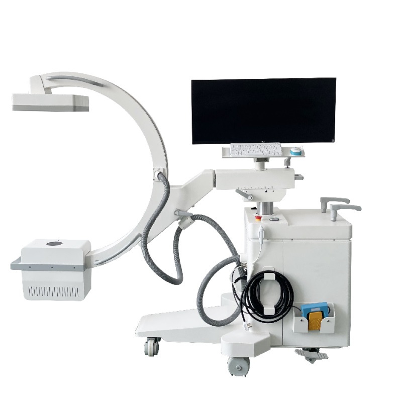
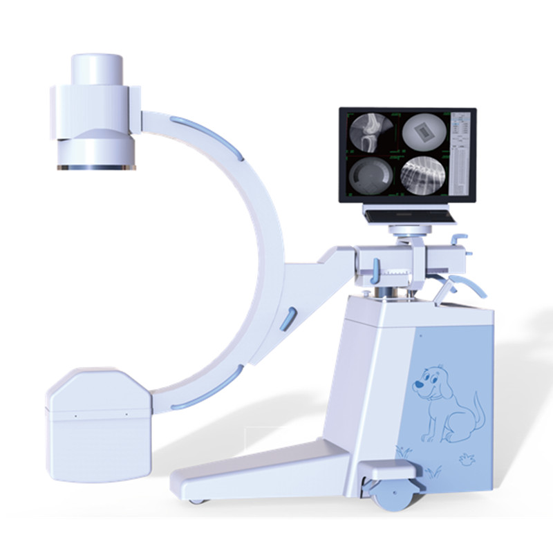
Powerful mobile C-arm machine designed for a wide range of applications in orthopedics, neurosurgery, pain management, and emergency room. With high frequency generator, it offers fine definition image with low dose. It brings to our users with unparalleled experience for operation when using the C-arm for orthopedic surgery and intervention operation.
High performance with excellent image quality thanks to a-Si detector. The 21×21cm image sensor has direct deposition CsI , which provides excellent low-dose imaging at frame rates up to 30 fps. When integrated into mobile C-arm fluoroscopic X-ray system, it is ideal for vascular or surgical applications.
Features
-With a compact appearance, and easy to operate.
-High -frequency generator emits high-quality and hard x-rays with strong penetrability.
-21cm*21cm flat panel detector, which has bigger image size than image intensifier.
-Amorphous silicon flat panel makes the surgery more accurate for the clinical operation due to high density resolution and spatial resolution.
-High stability oil-cooled heat-resistant combined tube, support long-time image acquisition in surgery, satisfy the requirement of long-time operation.
-34’’ monitor project real-time video and reference images simultaneously for quicker diagnosis and inference .
-Microcomputer control system with self-diagnostic function and automatic protection.
-Modular design offers error codes and reset button.
-Double CPU control improves the stability and safety of the equipment.
-Large opening design provides larger free diagnosis spaces, and maximizes the efficiency of surgery environment.
-Hand switch, foot switch and remote control make sure that you could do exposure inside and outside the operating theatre.
-PACS connectivity with DICOM 3.0 is convenient to send, receive and print images.
Technical Specifications
|
PARAMETERS |
SPECIFICATION |
|
X-ray generator |
|
|
Output power |
5KW |
|
Current of fluoroscopy |
0.3-6.3mA |
|
Current of radiography |
0.1-100mA |
|
Pulsed mA |
20mA |
|
Voltage |
40kV-125kV |
|
mAs range |
0.2-100mAs |
|
ms range |
10-1600ms |
|
X-ray tube |
|
|
Focal Spot value |
0.3mm /0.6mm |
|
Anode heat storage |
74KHU |
|
Housing Heat Storage Capacity |
1200KHU |
|
Tube continuous heat dissipation |
600W |
|
Flat panel detector |
|
|
Size |
21*21cm |
|
Detector Technology |
Amorphous Silicon |
|
Scintillator |
CsI |
|
Image resolution |
2.5lp/mm |
|
Frame rate |
MAX 30frames/sec |
|
Active pixels |
1024*1024 pixels |
|
A/D conversion |
16 bit |
|
Pixel Pitch |
205 μm |
|
Faster image capture speed |
The image will be displayed on the monitors in 0.8S after exposure. |
|
Grid |
|
|
Ratios |
10:1 |
|
Density |
60l/cm |
|
SID |
100cm |
|
C-arm unit |
|
|
SID |
1000mm |
|
Free space(Tube to I.I) |
820mm |
|
Arc depth |
640mm |
|
Vertical movement |
0-400mm(Electric) |
|
Horizontal movement |
0-200mm(Manual) |
|
Planning movement |
±15°(Manual) |
|
Orbital movement |
-30°-+90°(Manual) |
|
C-arc rotation |
±180° |
|
Workstation software |
|
|
Monitor |
One set 34’’ LCD HD monitor |
|
Computer memory |
500G mass storage. |
|
CPU |
2.7GHZ |
|
Software |
The image can be step-less rotated continuously for 360° |
|
Flip the Reference image horizontally or vertically |
|
|
Applies various Gamma values for each image |
|
|
Last frame frozen |
|
|
Images can be saved in different formats, such as JPG, DCM, RAW, etc. |
|
|
DICOM 3.0 |
|
|
Data communication |
|
|
Data storage etc. |
|


