Product Categories
-
Animal Monitoring & Life Support
-
Operating Theatre Equipment
-
Animal Diagnostic Laboratory
- Automatic Integrated Gene Detection System
- Hematology Analyzer
- Elisa Reader
- Chemistry Analyzer
- Blood Gas Electrolyte Analyzer
- Urine Analyzer
- Blood Gas&Immunoflurescence Analyzer
- Biological Safety Cabinet
- Biological Microscope
- Centrifuge
- Medical Pharmacy Refrigerator
- Blood Bank Refrigerator
- Combined Medical Refrigerator and Freezer
- Medical Freezer
- Laboratory Incubator
- Dry Oven
- Pet Rapid Test
-
Sterilizer (Autoclave)
-
Medical Imaging
-
Clinical Examination
-
Dental Equipment
-
Pet Grooming
product description
- Application
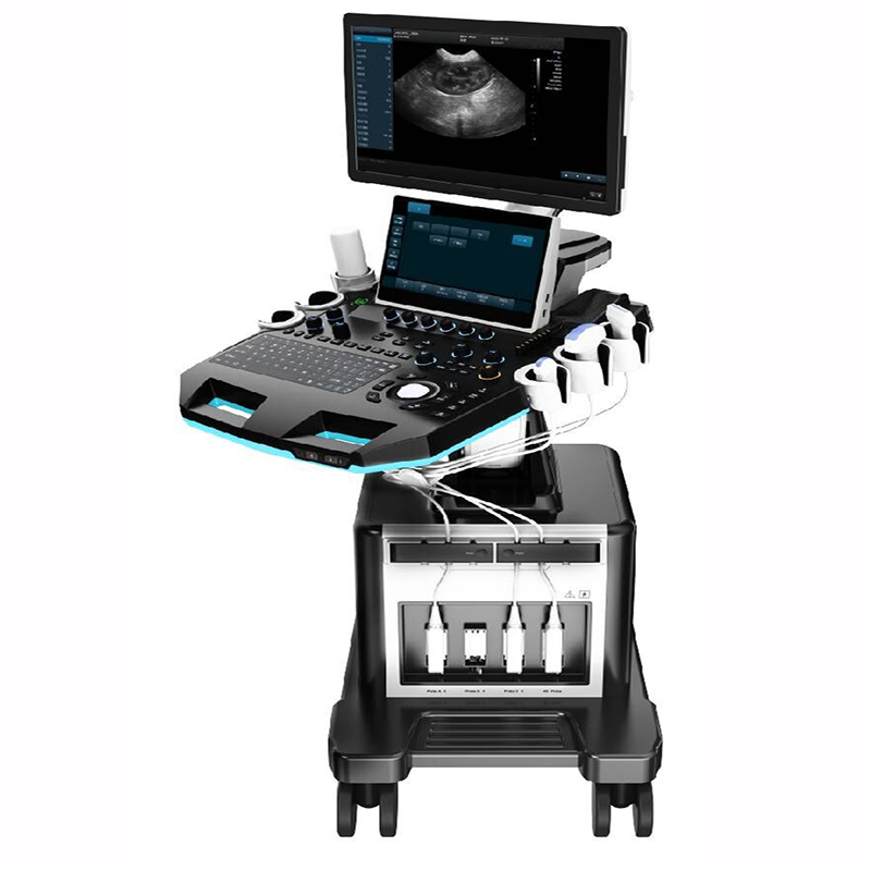
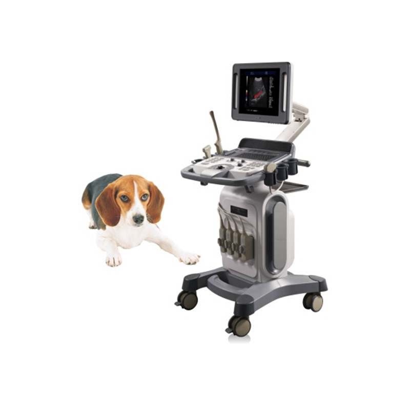
It’s applicable for animals’ examination and diagnosis on digestive system, reproductive system, urinary system for animal hospitals and scientific institutions.
- Technical Specifications
|
PARAMETER |
SPECIFICATION |
|
System technical specifications and summary |
|
|
Structure Style |
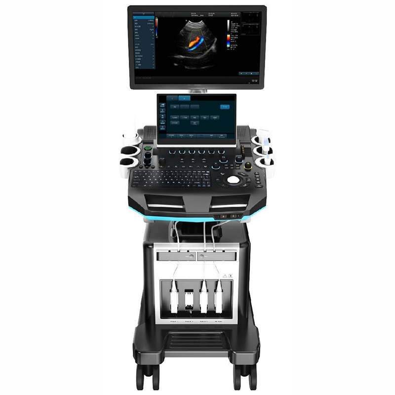
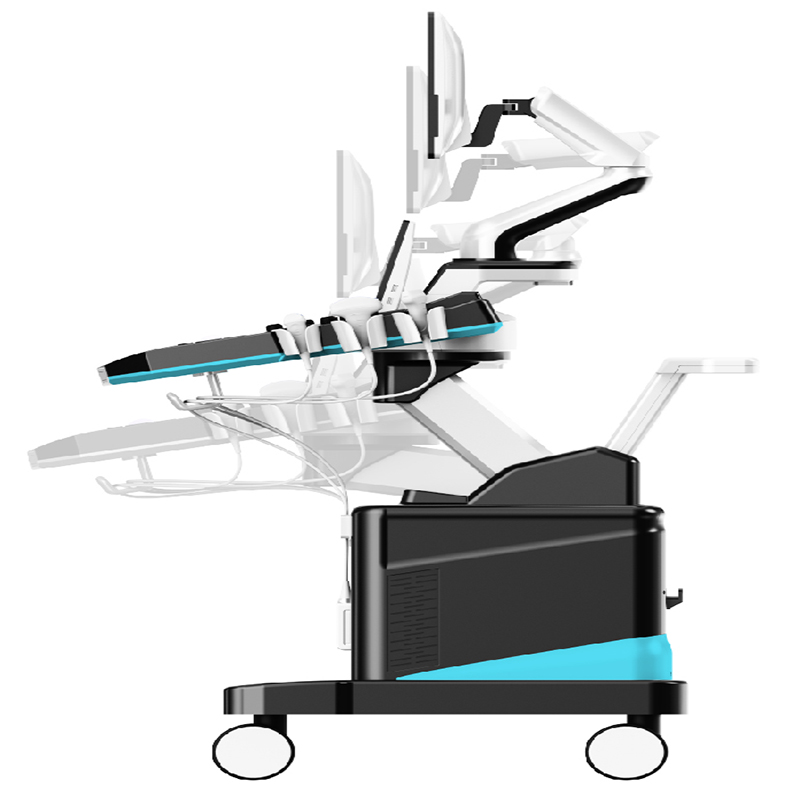
Dual Screen Cart |
|
Operating System |
Windows 10 |
|
Pulsed Wave Doppler Imaging (PW) |
|
|
Directional Power Doppler Imaging (DPDI) |
|
|
Real-time Triplex |
|
|
Spatial Compound Imaging |
|
|
Tissue Harmonic Imaging (THI) |
|
|
2B/4B Imaging Modes |
|
|
Support Language |
Chinese, English, French, Russian, Spanish |
|
Monitor sizes |
≥21.5 Inch |
|
All-in-one clipboard |
saved images display on the right of the screen, which can be directly transferred or deleted |
|
The system has the function of on-the-spot upgrade |
|
|
Preset table Conditions |
Preset optimal image inspection conditions to reduce adjustment during operation |
|
Support real-time 3D imaging |
|
|
Probe interface |
≥ 4 |
|
Trapezoidal Imaging |
|
|
One Key Smart Optimization |
|
|
Probe |
|
|
Convex probe |
Fundamental Frequency 2.5MHz/3.0MHz/3.5MHz/4.0MHz/H4.0MHz/H5.0MHz, six-segment frequency conversion (detecting depth: 30-255mm) |
|
Linear probe |
Fundamental Frequency 6.0MHz/7.5MHz/8.5MHz/10.0MHz/12.0MHz/H10.0MHz, six-segment frequency conversion (detecting depth 20-128mm) |
|
Phased probe |
Fundamental frequency 2.5MHz/3.0MHz/3.5MHz/4.0MHz/H3.0MHz/H4.0MHz.six-segment frequency conversion (detecting depth 100-244mm) |
|
Volume probe |
Fundamental frequency 2.0MHz/3.0MHz/4.5MHz/6.0MHz/H5.0MHz,five-segment frequency conversion (detecting depth 30-237mm) |
|
Micro-convex probe (R11) |
Fundamental frequency4.5MHz/6.0MHz/7.0MHz/9.0MHz/H8.0Mhz,five-segment frequency conversion (detecting depth 30-111mm) |
|
Rectal probe |
fundamental frequency 4.0MHz/6.5MHz/9.0MHz/H8.0MHz four-segment frequency conversion (detecting depth 20-110mm) |
|
High Frequency Phased Array |
Fundamental frequency 3.0MHz/5.0MHz/7.0MHz/H6.0MHz/H7.0MHz five-segment conversion (detecting depth 40-238mm) |
|
L25High Frequency linear probe |
Fundamental frequency 6.0MHz/7.5MHz/8.5MHz/10MHz/12MHz/H10.0MHz six-segment conversion (detecting depth 20-110mm) |
|
Backfat probe |
Fundamental frequency 2.0MHz/3.5MHz/4.0MHz/5.0MHz/H4.0MHz/H5.0MHz six-segment conversion (detecting depth 30-237mm) |
|
2D Imaging Mode |
|
|
Gain |
0-100 |
|
TGC |
8 segment adjustable |
|
Dynamic |
20-280, 20 levels adjustable |
|
Pseudo color |
0-11 level,visually adjustable |
|
Sound power |
Sound power: 5%-100%, step 5% visually adjustable |
|
Body mark |
≥18 kinds optional |
|
Maximum focus |
≥6, which can be moved throughout the whole process |
|
Grey scale map |
0-7, 7 levels adjustable |
|
Filter |
0-4 |
|
Scanning range |
50%-100% |
|
Frame correlation |
0-4, 4 level adjustable |
|
The screen has real-time display of sound power, probe frequency, dynamic range, pseudo color, gray scale and other 14 parameters can be adjusted |
|
|
Line density |
low, middle, high, 3 levels adjustable |
|
Noise reduction |
0-14 |
|
Noise reduction: 0-14 |
|
|
Color Frame correlation: 0-12, 12 levels visually adjustable |
|
|
Color map: 0-7, 7 levels visually adjustable |
|
|
Color flip: adjustable |
|
|
B / C real-time split screen mode |
|
|
Base line: 11 levels, visually adjustable |
|
|
Line density: low, high, 2 levels adjustable |
|
|
Filter: 0-5 levels adjustable |
|
|
Spectral Doppler Imaging Mode |
|
|
Sampling volume angle correction |
-80°~80°adjustable |
|
Sample volume |
0.5mm-20mm adjustable |
|
Frequency |
2.5MHz, 3.0MHz |
|
Base line |
9 levels, adjustable |
|
Pseudo color |
0-5 |
|
Parameter display |
≥4 kinds, adjustable |
|
Speed scale |
7.2-231cm/s (different probes have different ranges) |
|
Spectrum envelope function |
real time automatic spectrum envelope, manual spectrum envelope, and other. The system automatically analyses and displays various data such as PS, ED, RI, PI, S/D, HR, etc. |
|
Grey map |
0-7 |
|
Filter |
0-8 |
|
Dynamic range |
10-95db, step 5 |
|
Noise reduction |
0-28 |
|
Sound volume |
0-1007 |
|
3D imaging mode (optional) |
|
|
Quick Angle |
Support 0°, 90°, 180°, 270° rotation of the 3D window image |
|
Display layout |
Support “double””quad”, “single” image display |
|
Reconstruction mode |
Real-Skin, surface, Max, Min, XRax five reconstruction modes |
|
Pseudo-color display |
support 0-7 level adjustment |
|
Image magnification |
support level 5 |
|
Contrast |
0%-100% |
|
Threshold |
0%-100% |
|
Smooth |
≥3 levels adjustable |
|
X-axis, Y-axis, Z-axis rotation support adjustable |
|
|
Brightness |
0%-100% |
|
4D imaging mode (optional) |
|
|
Quick Angle |
Support 0°, 90°, 180°, 270° rotation of the 3D window image |
|
Display layout |
Support “double”, “quad”, “single” image display |
|
Reconstruction mode |
Real-Skin, surface, Max, Min, XRax five reconstruction modes |
|
Pseudo-color display |
support 0-7 level adjustment |
|
Image magnification |
support level 5 |
|
Contrast |
0%-100% |
|
Threshold |
0%-100% |
|
Smooth |
≥3 levels adjustable |
|
Pseudo-color |
≥7 levels adjustable |
|
X-axis, Y-axis, Z-axis rotation support adjustable |
|
|
Linear density |
support two-level adjustment |
|
Measurement and Analysis |
|
|
Measurement items include distance, area, angle, time, slope, heart rate, speed, acceleration, blood flow path, blood flow spectrum trace, resistance index/pulsatility index and other professional measurements |
|
|
Obstetrical measurement packages include dogs, cats, horses, cattle, and sheep |
|
|
Measurement line color and line type can be adjusted freely (including active color and finished color) |
|
|
The display position and font size of the measurement results can be adjusted as needed. |
|
|
Professional software package |
abdomen, obstetrics, urology, etc. |
|
Image and text management system: image saving format: BMP DCM JPG |
|
|
Host built-in ≥ 256G solid state hard disk starts quickly and stably |
|
|
Movie playback |
≥ 600 frames |
|
Built-in Chinese file information management system |
can record the number, name, inspection number, inspection date, etc., and can search and manage the number, inspection number, name, etc |
|
Report types ≥ 6. Provide photo proof. |
|
|
One-click quick report image and text management |
|
|
Interface |
|
|
USB ports, 1 Video, 1 S-Video, 1 DVI, 1 HDMI, 1 RJ-45. |
|
|
Configuration |
|
|
Color Doppler Ultrasonic Diagnosis System Host 1 |
|
|
Probe |
micro-convex R11 (optional), convex array probe (optional), linear array probe (optional), micro-convex R15 probe (optional), cardiac probe (optional), cavity probe (optional) , volume probe (optional), etc. |
|
Video printer (optional), laser printer (optional), etc. |
|


