Product Categories
-
Animal Monitoring & Life Support
-
Operating Theatre Equipment
-
Animal Diagnostic Laboratory
- Automatic Integrated Gene Detection System
- Hematology Analyzer
- Elisa Reader
- Chemistry Analyzer
- Blood Gas Electrolyte Analyzer
- Urine Analyzer
- Blood Gas&Immunoflurescence Analyzer
- Biological Safety Cabinet
- Biological Microscope
- Centrifuge
- Medical Pharmacy Refrigerator
- Blood Bank Refrigerator
- Combined Medical Refrigerator and Freezer
- Medical Freezer
- Laboratory Incubator
- Dry Oven
- Pet Rapid Test
-
Sterilizer (Autoclave)
-
Medical Imaging
-
Clinical Examination
-
Dental Equipment
-
Pet Grooming
product description
- Features
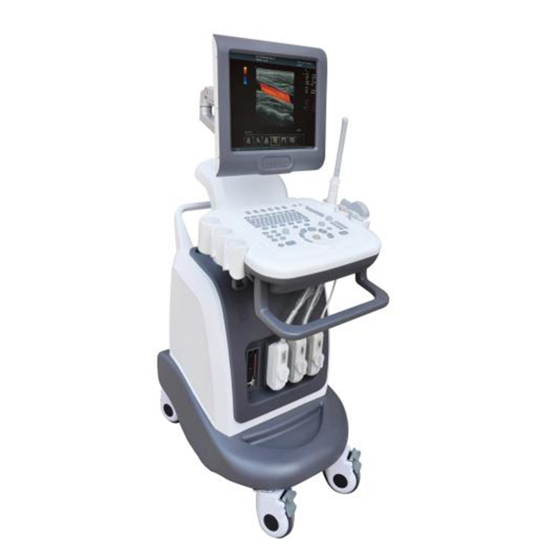
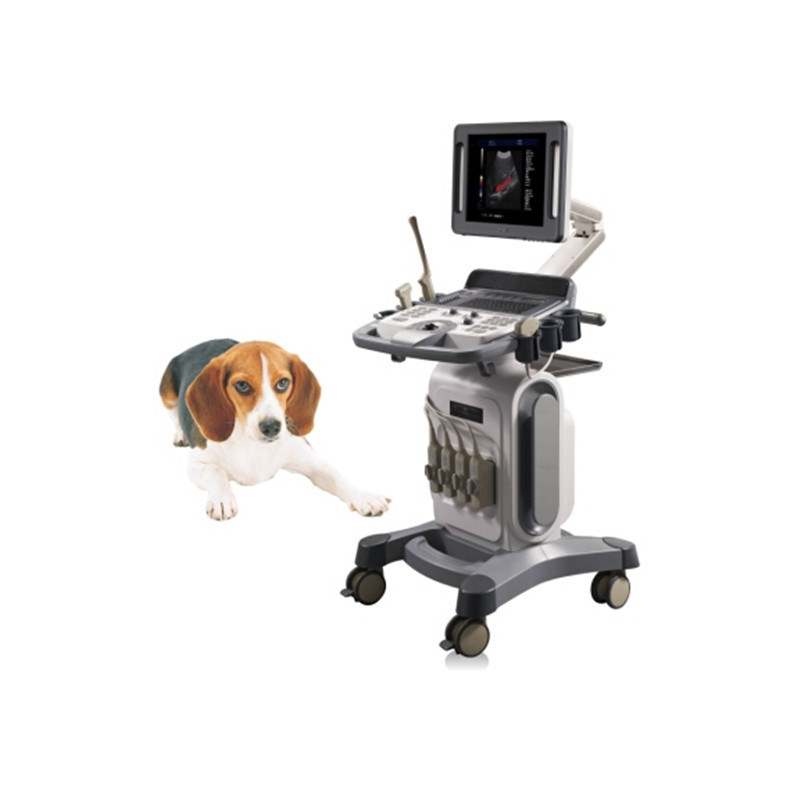
- Equipment use description: dog, cat, sheep, horse, cow, pig.
- Color Doppler flow imaging
- Two-dimensional gray scale imaging
- Spectrum Doppler display analysis system, PWD spectrum Doppler
- Energy Doppler imaging
- Tissue harmonic imaging
- With composite imaging, can be used for all probes, can be independently adjusted
- Image post-processing iclear function
- Technical Specifications
|
PARAMETER |
SPECIFICATION |
|
Measurement and analysis: |
General measurement: including distance, area, volume, time, heart rate, velocity, acceleration, Doppler routine, Doppler trace, etc Routine inspection report Custom comments: including insert, delete, edit, save, etc |
|
Input/output signal |
Output: S- video, USB, LAN |
|
Connectivity |
Medical digital imaging and communication DICOM3.0 interface components |
|
Image management and recording device |
hard disk, U disk storag |
|
Ultrasonic image archive and medical record management function |
complete the storage, management and playback of patient static image and dynamic image in the host |
|
Storage |
can be hard disk, U disk static and dynamic image storage |
|
Sound output safety |
The system has acoustic output power, mechanical index and thermal index display |
|
Color monitor |
15 inch high resolution color LCD monitor, no flicker, uninterrupted line by line scanning |
|
Probe interface |
2, 2 probe interface can be connected to all probes and interchangeable, not reserved other probe interface can not be universal, (non-external expansion port) |
|
Main parameters of two-dimensional gray scale imaging |
|
|
Maximum magnification of the image |
10 times |
|
Emission beam focusing |
4 focal points |
|
Reception mode |
multi-beam signal parallel processing |
|
The two-dimensional image gain adjustment range |
150dB, continuously adjustable |
|
maximum scanning depth |
30cm |
|
Beamforming device |
Digital beamforming device, digital whole-process dynamic focusing, digital dynamic variable aperture and dynamic trace, dynamic sideclobe compression, optimized transmission waveform, A/D≥12bit, focus position can be adjusted throughout the imaging area |
|
Playback |
the maximum playback of gray-scale images is 1000 pieces |
|
Preset conditions |
For different inspection organs, preset inspection conditions for optimized images, reduce adjustment during operation, and commonly required external adjustment and combination adjustment |
|
Gain adjustment |
B/M can be adjusted independently |
|
Sector scanning Angle |
10° -50 ° selection |
|
Visual adjustable dynamic range |
10-150dB |
|
STC segment adjustment 8 segments |
|
|
7 pseudo-color colors |
|
|
Clipboard image storage function in real-time diagnosis state |
|
|
Sound power 15 visual adjustable, step 1 |
|
|
Spectrum Doppler technical requirements |
|
|
Mode |
Pulse wave Doppler: PWD |
|
Doppler frequency |
linear array: PWD, 5 groups of frequencies; convex array: PWD, 5 groups of frequencies |
|
Sampling width and position range |
width 0.5-48mm |
|
The color filter has manual technology |
when the pulse repetition rate is adjusted, the wall filter can be optimized accordingly |
|
Display control |
reverse display (left/right; Up/down), B- Refresh |
|
Doppler envelope measurement and calculation |
|
|
Color Doppler technical requirements |
|
|
Doppler gain |
100%dB, continuously adjustable |
|
Display mode |
velocity variance display, energy display, velocity display, independent variance display, two-dimensional image/spectral Doppler/color blood flow imaging three synchronous display |
|
Display position adjustment |
The image range of interest in linear scanning: -10°- +10 |
|
Color Doppler chart quantitative analysis software |
blood flow velocity distribution map, blood flow measurement technology |
|
With the same screen left and right dual display B+COLOR function |
|
III. Probe specifications
- Optional probe group operating frequency range (2.0-10.0MHz)
- All probes have 8 groups of fundamental wave frequency and 8 groups of harmonic
frequency
- Harmonic imaging: can be applied to the configuration of convex array, linear
array, animal microconvex and animal rectal probes
- Abdominal convex array probe: operating frequency: 2.0-10.0MHz
- Linear probe: operating frequency: 2.0-10.0MHz
- Rectal probe: Operating frequency: 2.0-10.0MHz
- B/D dual-use: linear array: B/PWD, convex array: B/PWD, fan sweep: B/PWD
- Standard configuration
- Full digital color Doppler ultrasound host: 1 set;
- Instructions documents: 1 set
- Optional
- Rectal probe
- Convex probe
- Micro convex probe: 1 pc;
- Linear probe: 1 pc;


