Product Categories
-
Animal Monitoring & Life Support
-
Operating Theatre Equipment
-
Animal Diagnostic Laboratory
- Automatic Integrated Gene Detection System
- Hematology Analyzer
- Elisa Reader
- Chemistry Analyzer
- Blood Gas Electrolyte Analyzer
- Urine Analyzer
- Blood Gas&Immunoflurescence Analyzer
- Biological Safety Cabinet
- Biological Microscope
- Centrifuge
- Medical Pharmacy Refrigerator
- Blood Bank Refrigerator
- Combined Medical Refrigerator and Freezer
- Medical Freezer
- Laboratory Incubator
- Dry Oven
- Pet Rapid Test
-
Sterilizer (Autoclave)
-
Medical Imaging
-
Clinical Examination
-
Dental Equipment
-
Pet Grooming
product description
Features
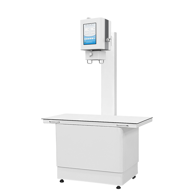
- Applicable to multiple scenarios, cost-effective choice.
- Removable, integrated integrated X-ray head, suitable for various use scenarios such as hospital and field.
- Three exposure modes: remote control, handle control and touch screen control, to meet the needs of different scenes.
- 17*17 inch amorphous silicon flat panel detector, clear imaging and clear layers.
- AED automatic exposure technology to ensure image quality, reduce scrap rate, and reduce repeated shooting.
- Bizcon image acquisition software with independent intellectual property rights, simple and intuitive workflow design, and practical measurement tools for medical specialists.
Technical Specifications
|
PARAMETER |
SPECIFICATION |
|
|
Fixed SID distance adjusted to 1000 mm |
||
|
Table Top Size |
1210 mm x 610 mm |
|
|
Whole Machine Dimension |
1200 mm x 860 mm x 2000 mm |
|
|
PARAMETER |
a-Si TFT Detector |
|
|
Pixel Size |
139μm |
|
|
Pixel Matrix |
2912 x 2912 |
|
|
AD Conversion |
16 bits |
|
|
Spatial Resolution |
3.6 lp/mm |
|
|
Imaging Area |
405mm × 405mm (17” x 17”) |
|
|
Dimension |
460mm x 460mm x 15mm |
|
|
Weight |
4Kg |
|
|
PARAMETER |
Portable X-ray Machine |
|
|
High Frequency |
40 KHz |
|
|
Single Phase |
220VAC+-10%, 5 KW |
|
|
Output Power |
100mA and 120Kvp |
|
|
X-ray tube H.U. continual monitoring for X-ray tube protection |
||
|
Kvp Radiographic range |
from 40 to 120 Kvp |
|
|
mA Radiographic range |
from 10 to 100 mA |
|
|
Anatomical program APR (Kvp, mAs, AEC, focal spot, etc) |
||
|
Light and sound indication for X-ray exposure |
||
|
Hand-Switch for preparation and exposure control. |
||
|
Manual collimator with 6 pair of leafs. |
||
|
Accessory rails (cones, filters, etc.) |
||
|
Field LED lamp of 160 LUX |
||
|
Light indicator for alignment with Bucky. |
||
|
Retractable measuring tape. |
||
|
Light indicator for alignment with Bucky. |
||
|
Retractable measuring tape |
||
|
PARAMETER |
Workstation and S/W (B-Console vet) |
|
|
Preview & Post processing Time : 3 sec |
||
|
KV, mA, mAS, Thumb nail View, patient Info and so on |
||
|
Main Screen Console: Veterinary patient information |
||
|
Automatic Pre & Post image enhancement. |
||
|
Expands image processing for optimized viewing |
||
|
Enhances variable density and form shading |
||
|
Variety Measurements |
||
|
Application measurements & teaching tools (such as VHS, HD, TPLO, TTA etc) |
||
|
One key to capture images into Clipboard |
||
|
DICOM sending and Report writing and Printing |
||
|
Easy Calibration & User setting |
||


